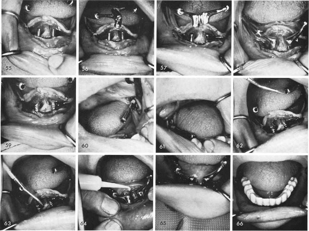
Both hollow tubes are seen, fig. 55. The anterior extensions are fitted into the tubes, figs. 56, 57, and the entire system is tapped downward until the anterior copings fit passively over the symphyseal blade posts, fig. 58. The anterior copings with their posterior extensions are released from the posterior hollow tubes by tapping the copings, extensions and hollow tubes in an up-ward direction, the blade posts notched and the hollow tubes are dried thoroughly, fig. 59. Figs. 60, 61 show the blades deeply into the rami and shows the exact area where the aluminous porcelain necks are located. The tubes and posts are moisturized with methyl methacrylate monomer, figs. 62, 63, and the polymer material is syringed into the hollow tubes and anterior copings to join the system together, fig. 64. Tissues are sutured anteriorly and in both posterior sections, fig. 65, and a temporary acrylic splint is cold cured, fig. 66. Healing is seen the day the sutures