| Mandibular Implants (published 1977) | Dr. Leonard I. Linkow |
|
|
Next Page |
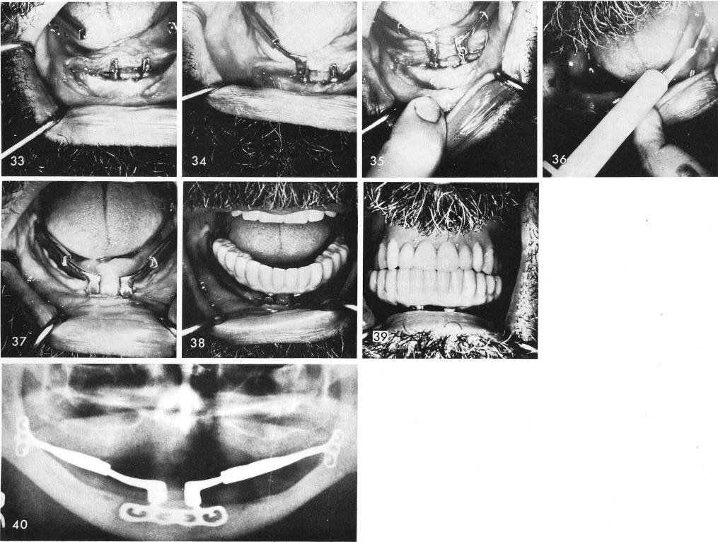
the right symphyseal blade post, fig. 34. It was repeated on the other side, fig. 35. With a syringe soft acrylic (Duralay) was syringed into the hollow tubes and anterior copings, fig. 36, and the system was joined together, fig. 37. Anteriorly, as shown here, horizontal transfixation pins were used to further lock the anterior copings. The acrylic splint, figs. 38, 39, and final x-ray, fig. 40.
304
|
|
Page 304 |
Next Page |
|
Copyright warning: This information is presented here for free for anyone to study online. We own exclusive internet copyrights on all content presented on this website. We use sophisticated technology to identify and legally close down websites that reproduce copyrighted content without permission - so please don’t do it.
|