| Mandibular Implants (published 1977) | Dr. Leonard I. Linkow |
|
|
Next Page |
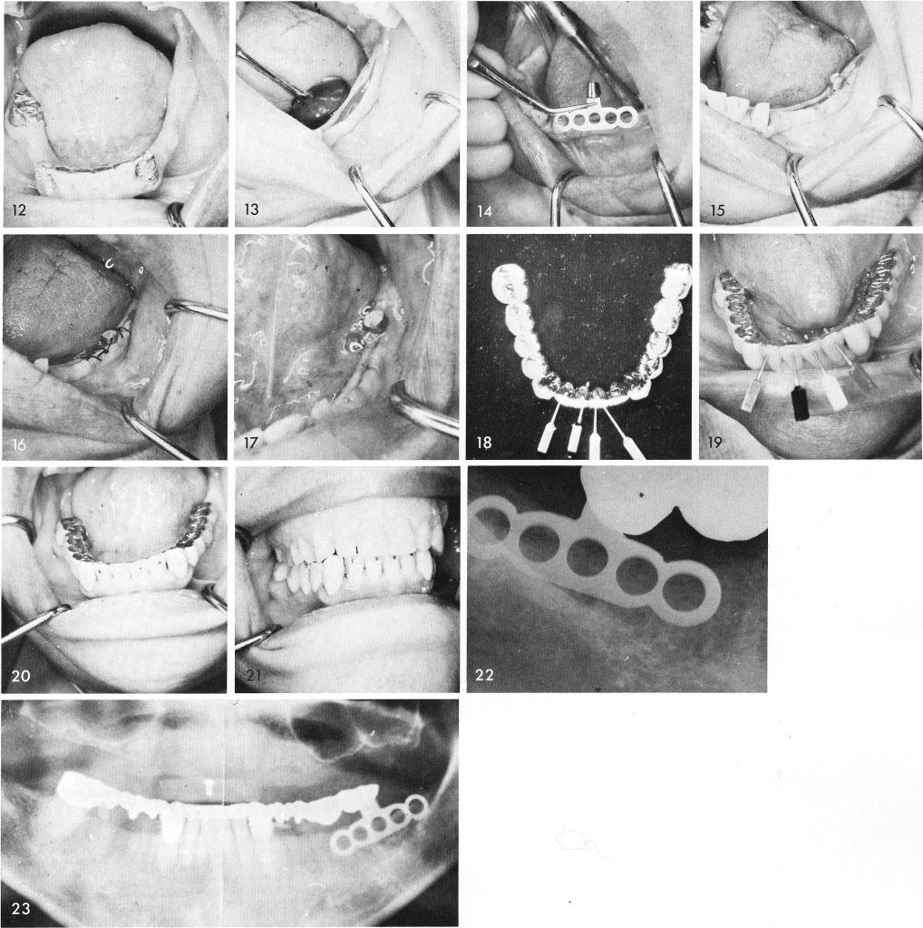
The tissues were then incised and reflected, fig. 13, exposing the knife-edge ridge and the earlier designed bladevent was inserted, figs. 14, 15, and sutured, fig. 16. After the sutures were removed, fig. 17, the acrylic veneer bridge was completed, fig. 18, and cemented into place with the non parallel pins cemented and "screwed" into position, figs. 19, 20, 21. Fig. 22 was the post-operative periapical film revealing the bladevent and fig. 23 shows a panoramic x-ray.
247
|
|
Page 247 |
Next Page |
|
Copyright warning: This information is presented here for free for anyone to study online. We own exclusive internet copyrights on all content presented on this website. We use sophisticated technology to identify and legally close down websites that reproduce copyrighted content without permission - so please don’t do it.
|