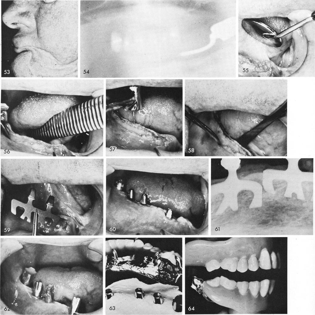
A patient with the entire left body and ramus of the mandible removed was helped considerably for several years before he deceased, fig. 53. A single molar tooth remained on his right side, fig. 54. The tissue was incised and reflected, figs. 55, 56, and a long groove was made along the entire length of the remaining half of the mandible, figs. 57, 58. Blades were carefully placed into the deepened socket, fig. 59. The tissues healed remarkably well, fig. 60. Notice the gold telescopic coping over the remaining molar tooth. Fig. 61 shows a periapical film revealing both blades. Copper bands festooned, shaped and filled with rubber material, fig. 62, were used in con-junction with a full arch rubber impression inside a tray to facilitate the laboratory procedures