| Theories and Techniques of Oral Implantology (vol.2) (published 1970) | Dr. Leonard I. Linkow |
|
|
Next Page |
| This is an archival HTML version of this book originally hosted here in 2006. The HTML may not display well on modern browsers. Please view the modern PDF Version for a better viewing experience. |
632 Theories and techniques of oral implantology
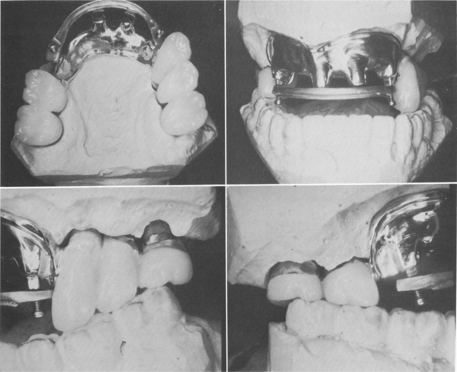
tachments were then cast in gold. After polishing, the template was soldered to the glazed porcelainfused-to-metal crowns, which had all been soldered to each other (Fig. 15-11).
A removable anterior quadrant consisting of individually waxed teeth and a pink saddle area con-forming to the anterior gold template contour was waxed (Fig. 15-12). It was fitted into the mouth, and all necessary adjustments were made.
A processed acrylic anterior quadrant of teeth with a pink acrylic portion resembling the patient's gums was then fabricated (Fig. 15-13). This was fused to the gold connecting bar that contained the
screws for attaching the anterior quadrant to the metal template.
The occlusion was once again checked and balanced in the mouth and the upper prosthesis re-moved. The two areas on the gingiva corresponding to the prefabricated hollow copings on the template were marked with indelible pencil. Because the alveolar bone in this area had resorbed to a knife-edge ridge and blind drilling seemed unwise, an incision was made to expose the bone. Then the implant sites were prepared (Fig. 15-14).
Two narrow ridge implants were carefully screwed into the ridge (Fig. 15-15) and positioned
B
D
A
C
Fig. 15-11. A, The posterior castings are soldered together and porcelain is glazed to them. Also seen is an anterior gold template soldered to the copings. B, The anterior view shows two anterior pontics, which are hollow inside to cover two implants that will be placed into the bone anteriorly, protruding from the template. The rounded bar is attached to the template by means of two screws seen. The bar will be processed into the anterior quadrant of acrylic teeth. C, Close-up view from the left side showing the three porcelain-fused-to-metal crowns soldered to the anterior metal template. A portion of the bar with the screw that fixates it to the template also is seen. D, Same view from the right side.
|
|
Page 632 |
Next Page |
|
Copyright warning: This information is presented here for free for anyone to study online. We own exclusive internet copyrights on all content presented on this website. We use sophisticated technology to identify and legally close down websites that reproduce copyrighted content without permission - so please don’t do it.
|