| Theories and Techniques of Oral Implantology (vol.2) (published 1970) | Dr. Leonard I. Linkow |
|
|
Next Page |
| This is an archival HTML version of this book originally hosted here in 2006. The HTML may not display well on modern browsers. Please view the modern PDF Version for a better viewing experience. |
Subperiosteal implants 573
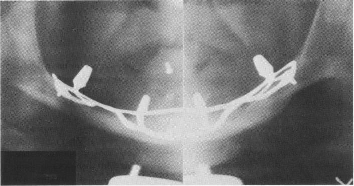
periosteal implant was made for the atrophied lower jaw (Fig. 12-110) .
Technique
An incision was made along the soft tissue covering the residual bony crest from tuberosity to tuberosity (Fig. 12-111, A). The tissue covering the hard palate was carefully retracted, exposing the posterior nasal spine and a portion of the greater and lesser palatine foramina (Fig. 12-111, B). An acrylic tray was fabricated directly over the hard palate (Fig. 12-111, C).
A rubber base impression was taken of the exposed palate using the acrylic tray for support (Fig. 12-111, D), and the tissues were sutured (Fig. 12-111, E) . A wax interocclusal record of centric relation was then taken.
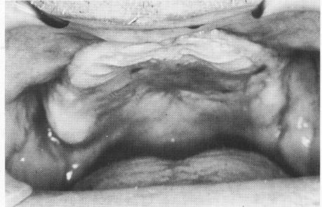
Fig. 12-109. The flattened maxillary ridge.
The implant was cast in cobalt-chrome. It contained two holes to accommodate two Vitallium screws to fix the implant to the hard palate. The casting also included three parallel posts 12 mm. long (Fig. 12-112, A). The implant was radiographed commercially to check for any porosities (Fig. 12-112, B) .
Two weeks later the tissue was again incised and retracted and the toroplant was screwed directly to the hard palate (Fig. 12-113). The tissues were then sutured. The three posts can be seen protruding through the central area of the soft tissues covering the hard palate (Fig. 12-114,A).
A hole was made in the bite registration to accommodate the protruding implant posts (Fig. 12-114 B and C) . The palatal portion of the tray was then relined with quick cure acrylic resin in order to obtain an accurate seat over the three posts (Fig. 12-114, D) . A wax interocclusal record of centric relation was taken.
The tissue healed uneventfully around the protruding implant posts in less than 10 days (Fig. 12-115 A). The tissue-bearing surface of the bite registration trays was lined with silicone material, and a final wax interocclusal record of centric relation was accomplished (Fig. 12-115, B).
A specially designed template was cast in gold. It contained two resilient anchorage attachments* that were only 1 mm. in height (Fig. 12-116). The circumferential attachments become an integral part of the denture.
*Rothermann's Resilience Anchorage-Eccentric 747, Cendres & Metaux, S. A. 2501, Biel-Biemme, Switzerland.
Fig. 12-110. A subperiosteal implant was fabricated for the mandible. Note the absence of bone below the sinus in the maxilla.
|
|
Page 573 |
Next Page |
|
Copyright warning: This information is presented here for free for anyone to study online. We own exclusive internet copyrights on all content presented on this website. We use sophisticated technology to identify and legally close down websites that reproduce copyrighted content without permission - so please don’t do it.
|