| Theories and Techniques of Oral Implantology (vol.2) (published 1970) | Dr. Leonard I. Linkow |
|
|
Next Page |
| This is an archival HTML version of this book originally hosted here in 2006. The HTML may not display well on modern browsers. Please view the modern PDF Version for a better viewing experience. |
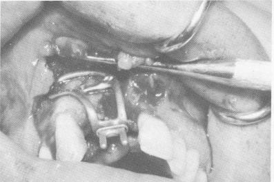
Fig. 12-92. The implant was fitted over the exposed bone.
Fig. 12-93. The implant was locked tightly to the bone when the lingual extensions of the transfixation pins were "riveted" to the lingual peripheral border of the substructure by grinding them flat with a heatless stone.
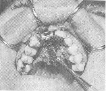
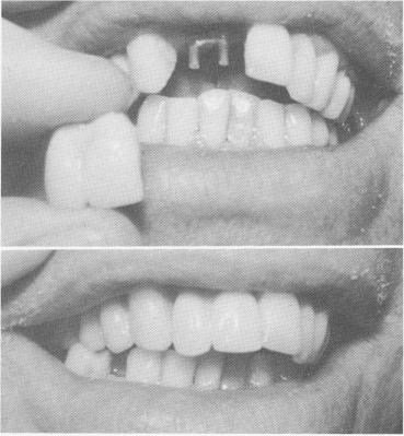
Fig. 12-94. The tissues were sutured to cover the implant and to closely adapt around the protruding post.
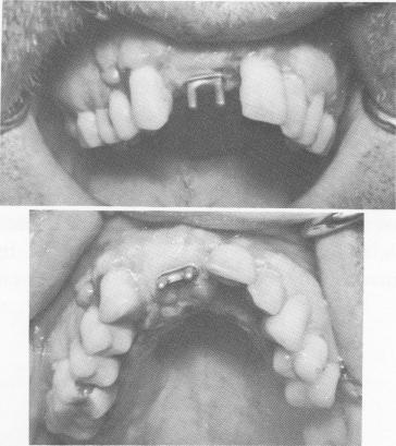
Fig. 12-95. After 3 weeks the tissue around the implant was completely healed.
Fig. 12-96. A two-unit acrylic splint was cemented over the protruding posts.
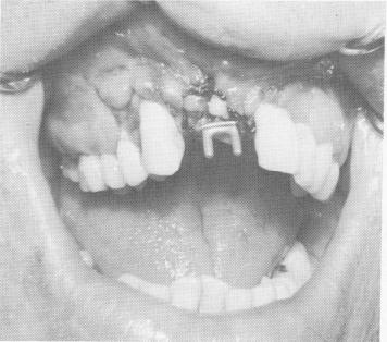
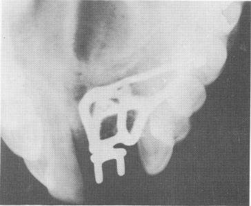
Fig. 12-97. A cross-sectional occlusal x-ray shows the subperiosteal implant with the horizontal transfixation pins.
|
|
Page 565 |
Next Page |
|
Copyright warning: This information is presented here for free for anyone to study online. We own exclusive internet copyrights on all content presented on this website. We use sophisticated technology to identify and legally close down websites that reproduce copyrighted content without permission - so please don’t do it.
|