| Theories and Techniques of Oral Implantology (vol.2) (published 1970) | Dr. Leonard I. Linkow |
|
|
Next Page |
| This is an archival HTML version of this book originally hosted here in 2006. The HTML may not display well on modern browsers. Please view the modern PDF Version for a better viewing experience. |
438 Theories and techniques of oral implantology
viouslv taken wax occlusal records and opposing jaw impressions the master stone model was articulated. A gold mesostructure consisting of smooth surfaced copings with high interproximal solder walls and veneer crown superstructures were cast (Fig. 10-192).
At the next visit, the copings comprising the mesostructure were fitted over the prepared abutment teeth (Fig. 10-193). Two internally threaded vent-plants were then screwed into the right and left maxillary central incisor regions until only about 1 mm. of their shafts protruded out of the fibromucosal tissue (Fig. 10-194). The veneer crown superstructures were then placed over the mesostructures (Fig. 10-195) and a wax interocclusal record of centric relation and a full mouth plaster index were taken( Fig. 10-196).
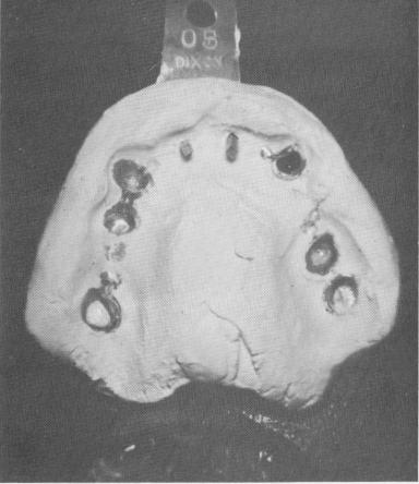
The master stone model was then made. Duplicate internally threaded implant posts were used as part of the mesostructure framework for the completion of the superstructure (Fig. 10-197). Obliquely-set channels were made in various strategically located portions of the mesostructure and superstructure wax-ups, and female internally threaded inserts were placed into the wax patterns before the wax frameworks were cast.
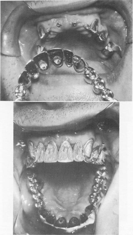
The full arch gold framework was tried in the mouth, and all necessary adjustments were accomplished (Fig. 10-198).
The gold was highly polished and the acrylic
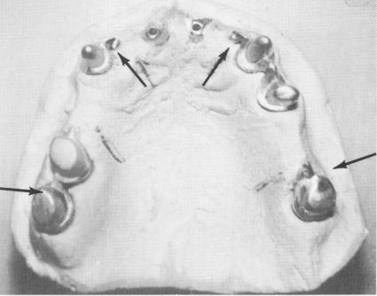
Fig. 10-196. A plaster index picks up the superstructure and mesostructure. Also seen are two duplicate implant shafts.
Fig. 10-197. The master stone model. Oblique channels were made at strategic areas in the mesostructure (arrows). (From Linkow, L. I.: Maxillary endosseous implants, Dent. Concepts 10[1 1:14-24, 1966.)
Fig. 10-198. The soldered full arch denture is tried in position.
|
|
Page 438 |
Next Page |
|
Copyright warning: This information is presented here for free for anyone to study online. We own exclusive internet copyrights on all content presented on this website. We use sophisticated technology to identify and legally close down websites that reproduce copyrighted content without permission - so please don’t do it.
|