| Theories and Techniques of Oral Implantology (vol.1) (published 1970) | Dr. Leonard I. Linkow |
|
|
Next Page |
| This is an archival HTML version of this book originally hosted here in 2006. The HTML may not display well on modern browsers. Please view the modern PDF Version for a better viewing experience. |
Implant histology 109
the fibrous tissue membrane separating the bone from the implant and thereby continued to receive their nourishment from this area.
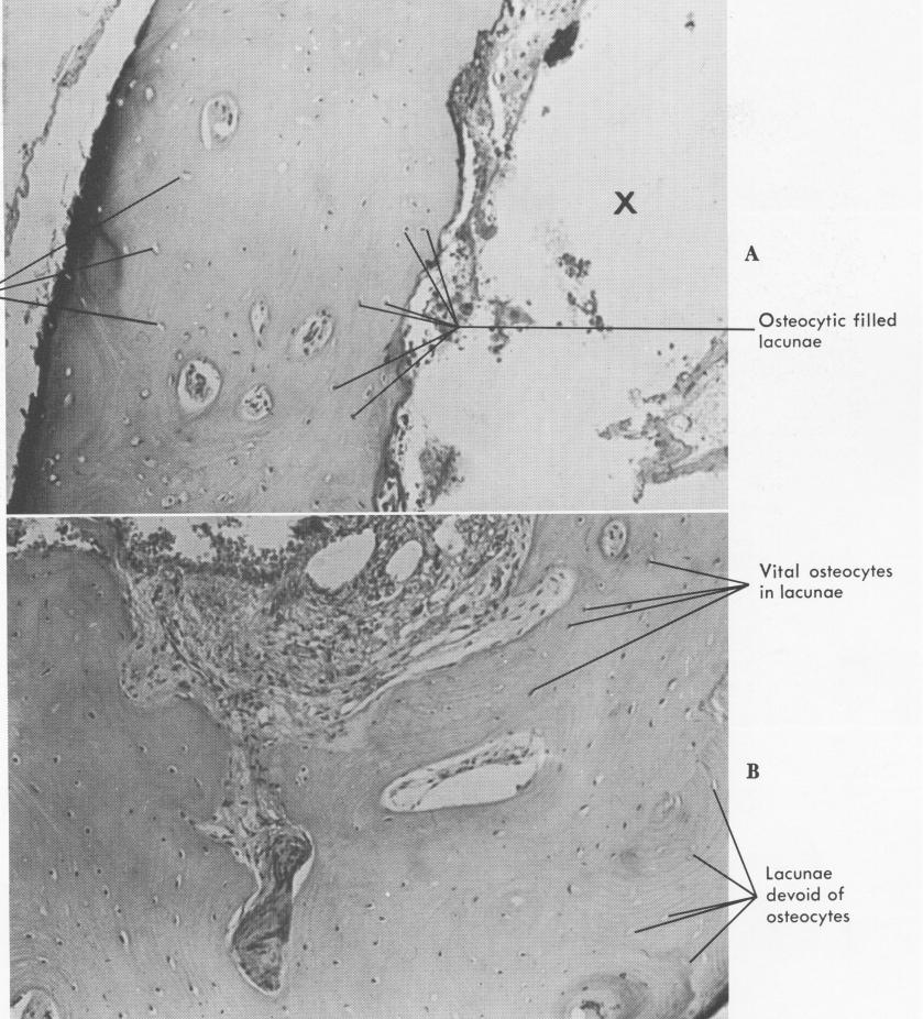
MICROSCOPIC DESCRIPTION: The specimen consists of a half-circle of compact bone with an associated smaller fragment [Fig. 4-54]. The con-figuration and size of bone is consistent with an area approximating the implant screw. The
outer aspect shows surface fragmentation and a narrow zone of basophilia. Lacunae in this area are devoid of osteocytes [Fig. 4-55]. The inner aspect of the specimen shows some foci of smaller chips of bone and pools of surgical hemorrhage [Fig. 4-56]. The bone along the inner aspect nearest the implant appears vital throughout [Fig. 4-57], showing numerous lacunae filled with osteocytes [Fig. 4-58]. The midportion of the bone dem-
Lacunae devoid of osteocytes
Fig. 4-55. A, The outer aspect of the bone has some empty lacunae, while the bone nearest the implant site appears vital. B, Another area of the same bone section, slightly enlarged. X, Implant site.
|
|
Page 109 |
Next Page |
|
Copyright warning: This information is presented here for free for anyone to study online. We own exclusive internet copyrights on all content presented on this website. We use sophisticated technology to identify and legally close down websites that reproduce copyrighted content without permission - so please don’t do it.
|