| Theories and Techniques of Oral Implantology (vol.1) (published 1970) | Dr. Leonard I. Linkow |
|
|
Next Page |
| This is an archival HTML version of this book originally hosted here in 2006. The HTML may not display well on modern browsers. Please view the modern PDF Version for a better viewing experience. |
112 Theories and techniques of oral implantology
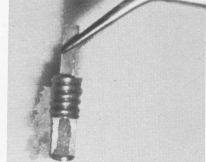
Fig. 4-61. The removed implant contains bone in its vent portion.
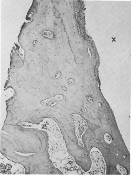
Fig. 4-63. A longitudinal section shows dense bone with numerous osteocytes around the implant site (X). (From Linkow, L. I.: Alloplastic implants. In Goldman, H. M., Forrest, S. P., Byrd, D. C., and McDonald, R. E.: Current therapy in dentistry, ed. 3, St. Louis, 1968, The C. V. Mosby Co.)
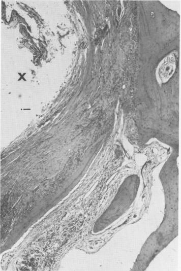
Fig. 4-62. The dense fibrous tissues around the implant site (X) is tightly bound to healthy bone, as seen in cross section. (From Linkow, L. I.: Alloplastic implants. In Gold-man, H. M., Forrest, S. P., Byrd, D. C., and McDonald, R. E.: Current therapy in dentistry, ed. 3, St. Louis, 1968, The C. V. Mosby Co.)
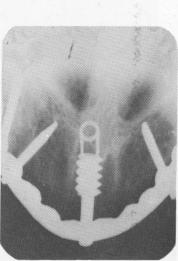
Fig. 4-64. This vent-plant broke 8 months after its insertion into the nasal septum.
|
|
Page 112 |
Next Page |
|
Copyright warning: This information is presented here for free for anyone to study online. We own exclusive internet copyrights on all content presented on this website. We use sophisticated technology to identify and legally close down websites that reproduce copyrighted content without permission - so please don’t do it.
|