| Theories and Techniques of Oral Implantology (vol.1) (published 1970) | Dr. Leonard I. Linkow |
|
|
Next Page |
| This is an archival HTML version of this book originally hosted here in 2006. The HTML may not display well on modern browsers. Please view the modern PDF Version for a better viewing experience. |
140 Theories and techniques of oral implantology
some bony repair was obvious in x-rays and there was no evidence of disease. Although the screw was moderately firm buccolingually, it could be rotated.
Today we can better explain, in light of modern experience, the implant's looseness. Basically, because the shape of the screw permitted the widest diameter to protrude into the mouth, a great deal of soft tissue invagination probably occurred. This invagination, together with the closeness of the screw's threads, did not allow the bone to grow close enough to the implant to follow its shape and tightly secure it.
The Strocks gathered more evidence on the fea
sibility of metallic implants through other experiments on humans. In the patient shown in Fig. 5-8, they inserted a Vitallium screw into the canine space of the right maxilla. Here failure was noticeable quite early. The implant was inserted into an open socket. The insertion was easy, and the implant quite loose. However, a celluloid crown was cemented into place and the screw left in to see if the bone would regrow around it anyway. At the same time, a maxillary left central incisor was extracted. After two weeks, the screw, which was in traurnatic occlusion, had become so loose that it had to be removed.
In the same patient, specially shaped Vitallium
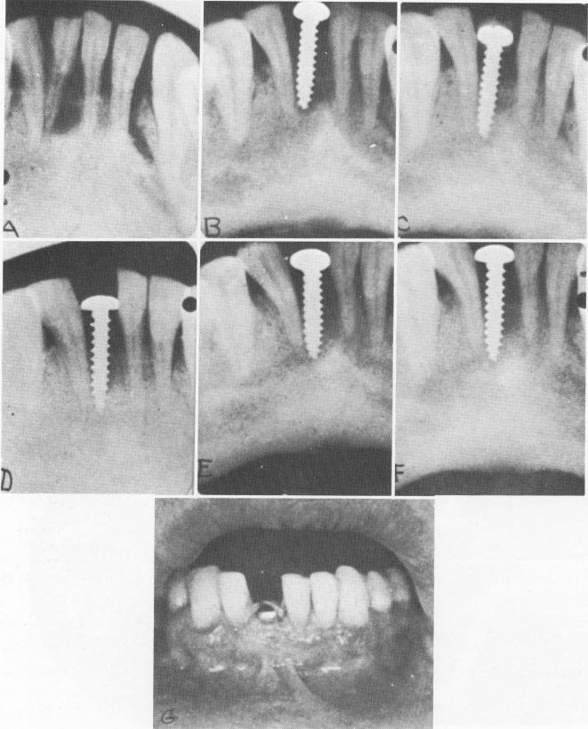
Fig. 5-7. A visual summary of a 1938 human implantation case done by the Strocks. A, The tooth to be extracted, with rarefaction about its apex. B, The implant, which was inserted in the socket about 3 weeks later. C through F, Bone condensation up to 8 months after insertion. G, The screw in position, showing the normal color and tone of the mucosa around the implant. (Courtesy A. E. Strock.)
|
|
Page 140 |
Next Page |
|
Copyright warning: This information is presented here for free for anyone to study online. We own exclusive internet copyrights on all content presented on this website. We use sophisticated technology to identify and legally close down websites that reproduce copyrighted content without permission - so please don’t do it.
|