| Theories and Techniques of Oral Implantology (vol.1) (published 1970) | Dr. Leonard I. Linkow |
|
|
Next Page |
| This is an archival HTML version of this book originally hosted here in 2006. The HTML may not display well on modern browsers. Please view the modern PDF Version for a better viewing experience. |
Operative tips 279
Miscellaneous instruments
Technologic innovations are facilitating implantology as well as other areas of specialization in dentistry and medicine. Sometimes the innovation is simple in both concept and construction. At other times the device is an outgrowth of several complex ideas. Whether or not the operator need avail himself of such a device, which generally tends to be expensive, usually depends on the amount of use he can derive from it. Linkow has found the following instruments useful, but not necessary, adjuncts to his implantation procedures.
X-ray grid. The x-ray grid is a mesh with 1 sq. mm. spaces used in conjunction with x-rays (Fig. 7-53). The grid enables an operator who is unsure of the amount of bone remaining in the alveolar crest to accurately measure it in millimeters (Fig. 7-54). From the reading he can exactly choose an implant of the appropriate design and length and mark the ideal placement depth on the implant itself. While drilling he can keep accurate track of
the amount of bone relative to instrument length (Fig. 7-55).
To superimpose the grid onto the radiograph, the grid is taped to the tongue side of the x-ray fihn itself prior to exposing it. Before development of the film, the grid is removed and reserved for other use.
Self-contained developer and fixer for dental x-ray films. For a quick chairside developing and fixing of an x-ray without the use of a darkroom, a self-contained x-ray film unit can be used (Fig. 7-56). The film is included in a black plastic casing. The film compartment is joined to a narrow chamber that leads to two separate chambers that contain developer and fixer.
After the x-ray is exposed, the developer chamber is opened by pulling the single tail that extends from it. The developing solution is squeezed from its compartment through the narrow chamber and into the x-ray film department. After 1 or 2 minutes, the fixer solution is opened by pulling its two
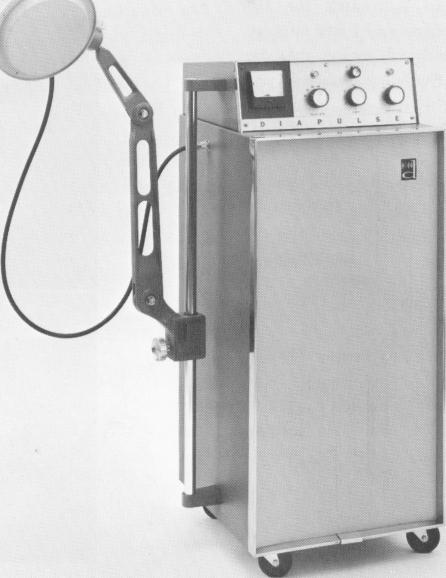
Fig. 7-57. A Diapulse machine. (Courtesy Diapulse Corporation of America.)
|
|
Page 279 |
Next Page |
|
Copyright warning: This information is presented here for free for anyone to study online. We own exclusive internet copyrights on all content presented on this website. We use sophisticated technology to identify and legally close down websites that reproduce copyrighted content without permission - so please don’t do it.
|