| Theories and Techniques of Oral Implantology (vol.1) (published 1970) | Dr. Leonard I. Linkow |
|
|
Next Page |
| This is an archival HTML version of this book originally hosted here in 2006. The HTML may not display well on modern browsers. Please view the modern PDF Version for a better viewing experience. |
306 Theories and techniques of oral implantology
central incisor pontic was prepared for the full crown preparation (Fig. 7-93). A vent-plant was then screwed into the upper right cuspid region (Fig. 7-94). An elastic impression was taken, including the posterior edentulous area, the protruding implant post, the right central incisor pontic, and the entire left side of the arch. A wax interocclusal record of centric relation was taken, as well as a lower elastic impression from which the final fixed partial denture was fabricated (Fig. 7-95). The bridge was cemented over the central incisor pontic preparation and the cuspid vent-plant, and pin implants were drilled through the scalloped template in the posterior edentulous area (Fig. 7-96). The pins were fused with acrylic (Fig. 7-97) and the molar crown cemented over the core (Fig. 7-98). The completed unilateral fixed partial denture thus became part of the full arch splint (Fig. 7-99).
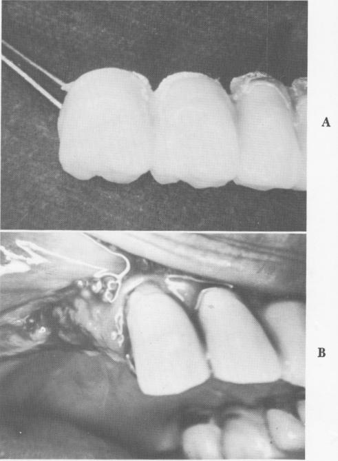
Many times a new fixed partial denture can be fabricated immediately after the loose pins are re-moved, as in the following case. The bridge was severed at the solder joint distal to the cuspid crown (Fig. 7-100). The bridge was easily pulled out with the pins still attached (Fig. 7-101). The porcelain was cut away from the cuspid crown and the under-lying gold prepared for a full coverage crown, making sure not to separate the solder joint on its mesial aspect from the lateral incisor. The prefabricated unilateral fixed partial denture was processed (Fig. 7-102) and cemented in position (Fig. 7-103). Pin implants were then drilled through the predetermined holes in the template, which was angled to circumvent the sinus (Fig. 7-104). The pins were joined together with cold cure acrylic and the super-structure cemented over the acrylic core and gold post (Fig. 7-105).
Although it may be easy in most cases to remove a failing triplant still attached to the restoration, in those cases where the pins do not easily slip out after severing the crowns other measures should be taken to avoid further damage to the bone. The crown should be separated from the acrylic core and the core disassembled by cutting into it with either a fissure bur, large round bur, or vulcanite bur. When the pins have been exposed, each is removed individually using a narrow-pointed pliers (Fig. 7-106).
In cases where one pin may have perforated the labial or buccal plate of bone, it may be possible to save the implant by shortening the offending pin. The perforated area is determined by palpation and radiographs, and then the soft tissues are incised and reflected to expose the protruding portion.
Fig. 7-101. A, The pin implants are removed along with the bridge. B, The site immediately after removal of the bridge.
The offending part is cut off flush with the bone (Fig. 7-107) and the tissues closed. Providing that the pins have been correctly angled, the shortened pin should not endanger the triplant's stability (Fig. 7-108).
If the offending pin must be entirely removed, it is gripped with a narrow-pointed pliers—preferably a Howe pliers and pulled out in the same direction along which it was angled. If there is too much resistance from the acrylic core, then it may be necessary to remove the entire implant by involving the entire acrylic core, its overlying crown, and neighboring solder joints. In most cases, however, the prostheses can usually be maintained with a bit of ingenuity.
Failing blade implants
Blade implants, because of their unique design and extremely large openings, are more difficult to remove than the post and pin type implants. Even those blades that are failing because of poor tech-
|
|
Page 306 |
Next Page |
|
Copyright warning: This information is presented here for free for anyone to study online. We own exclusive internet copyrights on all content presented on this website. We use sophisticated technology to identify and legally close down websites that reproduce copyrighted content without permission - so please don’t do it.
|