| Theories and Techniques of Oral Implantology (vol.1) (published 1970) | Dr. Leonard I. Linkow |
|
|
Next Page |
| This is an archival HTML version of this book originally hosted here in 2006. The HTML may not display well on modern browsers. Please view the modern PDF Version for a better viewing experience. |
330 Theories and techniques of oral implantology
full crown restoration, the gingival tissue was retracted, and an elastic impression was taken to include the prepared tooth, the edentulous area anterior to it, and the cuspid tooth. A wax bite was also taken. A two-unit acrylic-and-gold-veneer pros-thesis was processed (Fig. 8-62). It was tried in the mouth and all necessary adjustments were made. A specially sized helical bur was then used in a contra-angle to create a hole in the desired area of bone (Fig. 8-63). An appropriate narrow ridge im-
Fig. 8-68. The splint is carefully balanced.
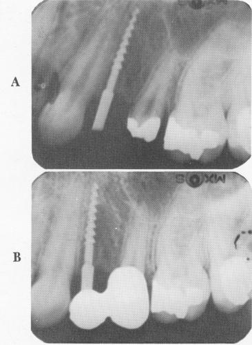
Fig. 8-69. A, An intraoral radiograph showing the narrow ridge implant in the bone to its desired depth. B, The x-ray shows the prosthesis cemented over the implant and tooth abutment.
plant was selected (Fig. 8-64), inserted into the hexagonal prolongator (Fig. 8-65), and carefully screwed into place (Fig. 8-66).
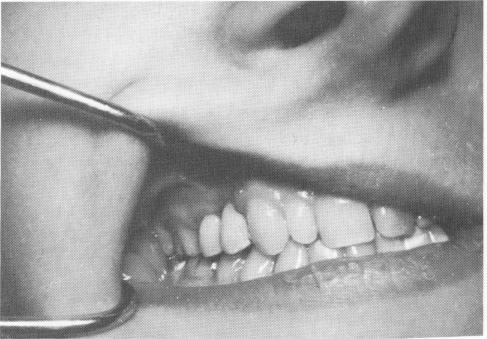
The two-unit, prefabricated splint was fitted over the implant shaft and prepared bicuspid tooth (Fig. 8-67). (If any interference occurs from such a post, a larger hole can be made on the under-surface of the abutment crown.) The bridge was then cemented into position with a hard cement (Fig. 8-68). During this procedure, as should be done during all implant interventions, x-rays were taken as guides (Fig. 8-69).
UNUSUAL SINGLE TOOTH IMPLANTS
A significant number of presenting cases raise atypical problems. The following are characterized
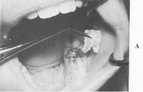
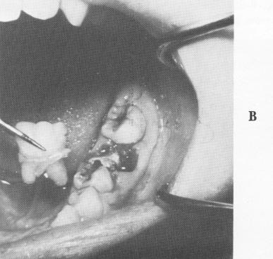
Fig. 8-70. A, The internal surface of the buccal half of a completely sagittally fractured mandibular first molar. B, Another view showing the buccal surface of the crown and roots. The lingual half of the tooth is still in the bone.
|
|
Page 330 |
Next Page |
|
Copyright warning: This information is presented here for free for anyone to study online. We own exclusive internet copyrights on all content presented on this website. We use sophisticated technology to identify and legally close down websites that reproduce copyrighted content without permission - so please don’t do it.
|