| Theories and Techniques of Oral Implantology (vol.1) (published 1970) | Dr. Leonard I. Linkow |
|
|
Next Page |
| This is an archival HTML version of this book originally hosted here in 2006. The HTML may not display well on modern browsers. Please view the modern PDF Version for a better viewing experience. |
90 Theories and techniques of oral implantology
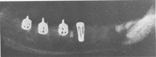
Fig. 4-14. Pasqualini experimentally buried several implant designs in dogs' jaws. (From Pasqualini, U.: Reperti anatomopatalogici e deduzioni clinico chirurgiche di 91 impianti alloplastici in 28 animali da esperimento, Riv. Ital. Stomat., No. 12, December, 1962.)
is not uncommon; however, the trabeculae are never in contact with metal, and numerous interpretations of their presence may be proposed.
Photomicrographs of the site revealed fibrous tissues with an osseous portion on the crest border [Fig. 4-8], young connective tissues with a pre-dominance of histiocytes [Fig. 4-9], and superficial epithelium with malpighian bodies [Fig. 4-10]. Also evident were fibrous tissues consisting of fibrocytes with some plasmodial elements [Fig. 4-11], fibrous tissues very rich in cellular elements with plasmodial elements [Fig. 4-12], and fibrocytic connective tissues [Fig. 4-13].
Perron-Andres concluded from the various sections taken from the site that had borne an implant for 6 months that:
-
The implant did not promote the kinds of tissue reactions associated with a foreign body inclusion.
-
The epithelial lining, although it invaginated down to the penultimate spiral, was prevented from completely encircling the implant and exfoliating it by the dense connective tissues and bone at the apex of the implant.
-
The differentiation of connective tissue to-ward the organization of a proteolytic web is variable.
-
Slowly organizing connective tissue surrounds the apical spiral and holds the implant steady, permitting normal mastication. However, the implant is slightly more mobile than a natural tooth.
-
Reactions of the tissues around an implant can vary from the formation of connective fibrous tissues with poor osseous differentiation to dense bone reconstruction. As long as the type of tissue is not too excessive in either direction, the implant can be expected to function normally.
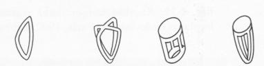
Fig. 4-15. Other Pasqualini implants, showing their methods of insertion. (From Pasqualini, U.: Reperti anatomopatalogici e deduzioni clinico chirurgiche di 91 impianti alloplastici in 28 animali da esperimento, Riv. Ital. Stomat., No. 12, December, 1962.)
Seidenberg and Lord's buried thimbles
In 1963, Seidenberg and Lord reported on their insertion of Vitallium thimbles into the fresh molar sockets of five mongrel dogs. Ten weeks after insertion histologic sections revealed a very thin collagenized band of connective tissue adjacent to an implant, with the remainder of the socket composed of coarse cancellous bone.
Seidenberg and Lord concluded that by 10 weeks the healing process was essentially complete insofar as immobilizing the implant was concerned. How-ever, they suggested that further maturation of bone might be encouraged prior to subjecting the implants to stress. Other investigators, Linkow included, have since found that an implant placed in immediate function actually helps the bone mature by restoring near-natural stress on it.
|
|
Page 90 |
Next Page |
|
Copyright warning: This information is presented here for free for anyone to study online. We own exclusive internet copyrights on all content presented on this website. We use sophisticated technology to identify and legally close down websites that reproduce copyrighted content without permission - so please don’t do it.
|