| Mandibular Implants (published 1977) | Dr. Leonard I. Linkow |
|
|
Next Page |
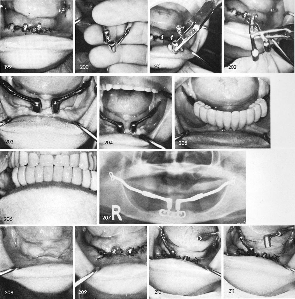
recast. Fig. 199 shows the tissues 24 hours later and the posterior horizontal extensions after the anterior portion was cut loose. The newly cast anterior copings with posterior horizontal tubes, figs. 200, 201, 202. The healed case, figs. 203, 204, and the teeth, figs. 205, 206, and post-operative x-ray, fig. 207.
Another case where a subperiosteal failed because of posterior tissue problems on both sides, fig. 208. The ramus blade is inserted and the tissues are sutured, fig. 209. The procedure continued to join anterior and posterior portions together, figs. 210, 211, 212. The healing was excellent, fig. 213, and the case completed, figs. 214, 215, 216, and the final x-ray, fig. 217.
317
|
|
Page 317 |
Next Page |
|
Copyright warning: This information is presented here for free for anyone to study online. We own exclusive internet copyrights on all content presented on this website. We use sophisticated technology to identify and legally close down websites that reproduce copyrighted content without permission - so please don’t do it.
|