| Mandibular Implants (published 1977) | Dr. Leonard I. Linkow |
|
|
Next Page |
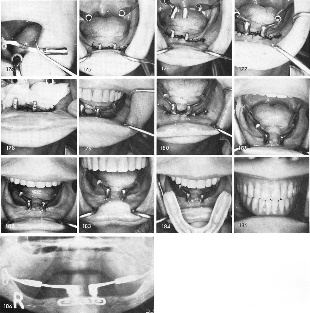
posts, figs. 176, 177, then removed so the various parts could be dried thoroughly, fig. 178, and cemented into position, figs. 179, 180. Figs. 181, 182, 183 reveals the healing.
The denture is adjusted and cemented with Duralay, figs. 184, 185. Fig. 186 shows the post-operative x-ray.
315
|
|
Page 315 |
Next Page |
|
Copyright warning: This information is presented here for free for anyone to study online. We own exclusive internet copyrights on all content presented on this website. We use sophisticated technology to identify and legally close down websites that reproduce copyrighted content without permission - so please don’t do it.
|