| Mandibular Implants (published 1977) | Dr. Leonard I. Linkow |
|
|
Next Page |
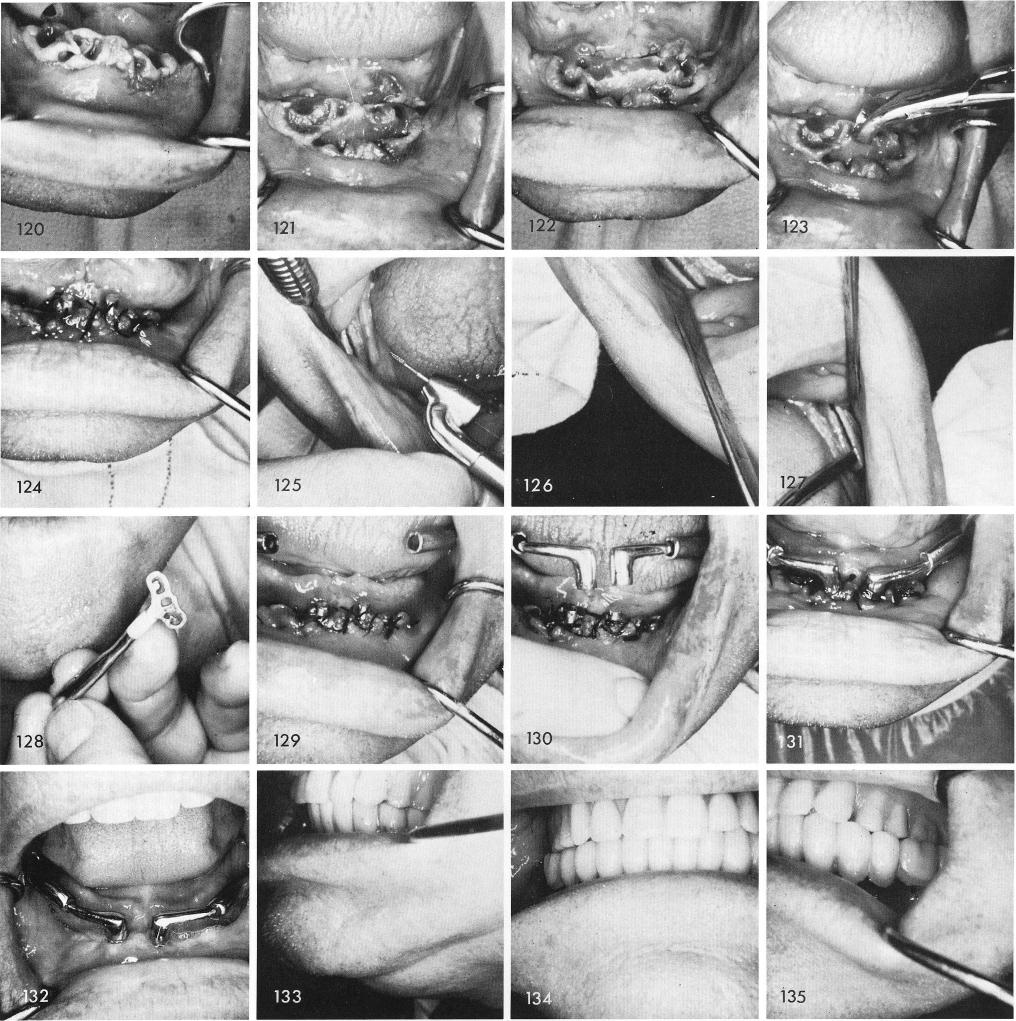
the bone exposed, and ridge widened to accept the symphyseal bladevents, figs. 121, 122, 123, 124. The groove was made in the right ramus, figs. 125, 126, and the left ramus, fig. 127. The blades were inserted, figs. 128, 129, and the anterior copings with their extensions were fitted, cemented and locked into place, figs. 130, 131, and the tissues were sutured closed. The healed site and temporary lower splint, figs. 132, 133. Figs. 134, 135, 136 show the preliminary wax-up.
311
|
|
Page 311 |
Next Page |
|
Copyright warning: This information is presented here for free for anyone to study online. We own exclusive internet copyrights on all content presented on this website. We use sophisticated technology to identify and legally close down websites that reproduce copyrighted content without permission - so please don’t do it.
|