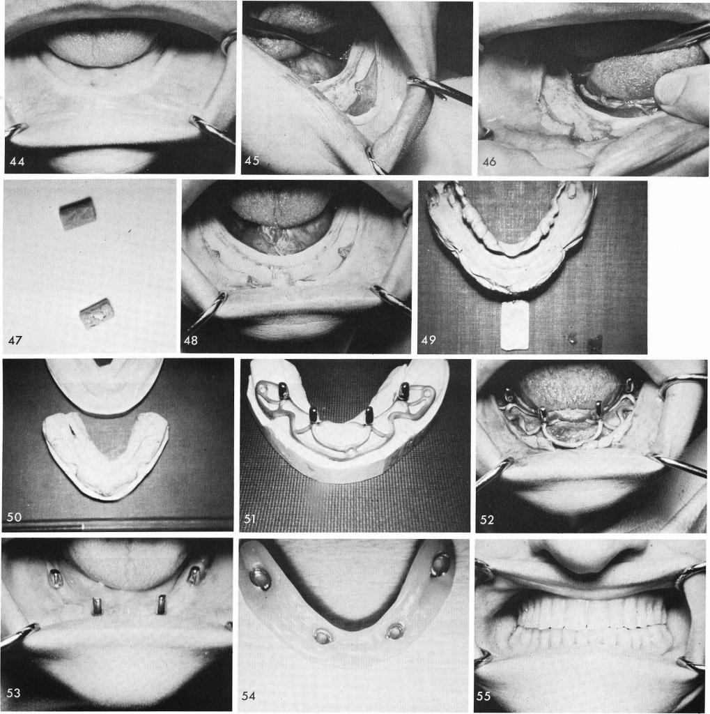
The patient accepted a subperiosteal implant and procedures were started, fig. 44. The bone was exposed, exposing the both mental bundles and the areas where the magnets were implanted posteriorly, figs. 45, 46. The magnets were removed, fig. 47, the bone cleansed of soft tissue debris, fig. 48, and an accurate rubber base impression was taken. Notice the impression included the areas where the magnets were removed, fig. 49. Fig. 50 shows stone models revealing the difference in contour between the soft tissue impression and bone impression. The implant, fig. 51, is fitted into place, fig. 52. Healing was good, fig. 53. The processed denture was then seated, figs. 54, 55.