| Mandibular Implants (published 1977) | Dr. Leonard I. Linkow |
|
|
Next Page |
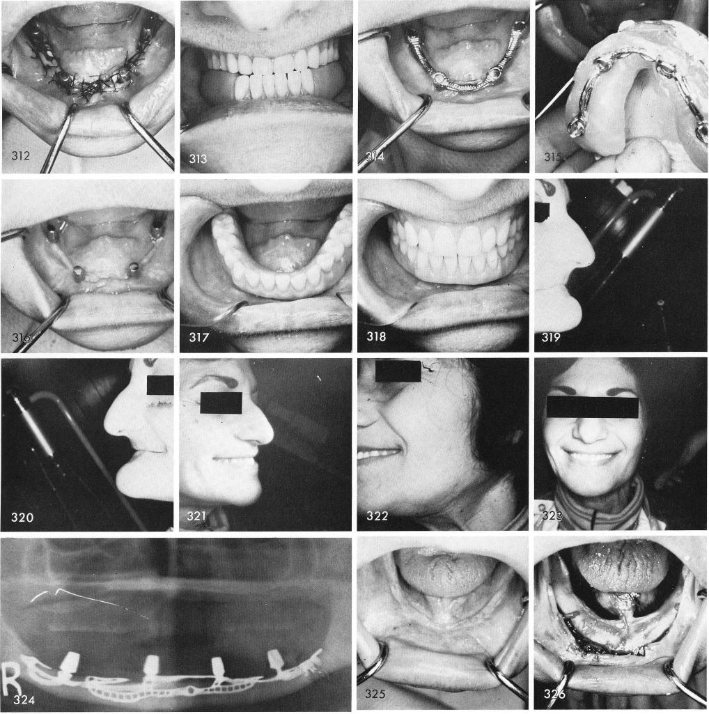
was made, figs. 308, 309. Figs. 310, 311 shows the nerves to be intact in the newly prepared canals. The tissues were carefully sutured being certain not to suture too deeply thus involving the nerve bundles, fig. 312. The temporary stent, fig. 313. The tissue healed excellently, fig. 314, a wax bite was taken, fig. 315. Six weeks later shows good tissue healing, fig. 316. The denture and subperiosteal implant have been successfully functioning for over seven years, figs. 317, 318. Before and after shots, figs. 319, 320, 321, 322, 323. The post-operative x-ray, fig. 324.
Another severely resorbed mandible, fig. 325, showing the extreme shallowness of the bony structure, fig. 326. An impression of heavy and light Citricon was taken, fig. 327, and the tissues sutured together, fig. 328.
280
|
|
Page 280 |
Next Page |
|
Copyright warning: This information is presented here for free for anyone to study online. We own exclusive internet copyrights on all content presented on this website. We use sophisticated technology to identify and legally close down websites that reproduce copyrighted content without permission - so please don’t do it.
|