| Mandibular Implants (published 1977) | Dr. Leonard I. Linkow |
|
|
Next Page |
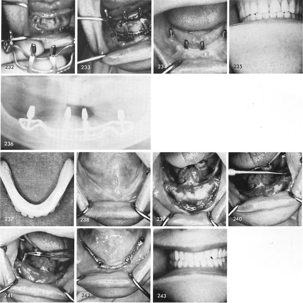
fitted over the bone accurately and the anterior cross-over struts fitted exactly into the grooves, figs. 232, 233. Healing was excellent, fig. 234, and the dentures given to the patient, fig. 235. Fig. 236 is the post-operative x-ray.
A convex tissue bearing denture surface, fig. 237, revealing a concave clinical picture, fig. 238. Enough bone still existed anteriorly, fig. 239, although the inferior alveolar nerve was easily lifted out of its unprotected canal, figs. 240, 241. Healing, here too, was excellent, fig. 242, and the case successful, fig. 243.
273
|
|
Page 273 |
Next Page |
|
Copyright warning: This information is presented here for free for anyone to study online. We own exclusive internet copyrights on all content presented on this website. We use sophisticated technology to identify and legally close down websites that reproduce copyrighted content without permission - so please don’t do it.
|