| Mandibular Implants (published 1977) | Dr. Leonard I. Linkow |
|
|
Next Page |
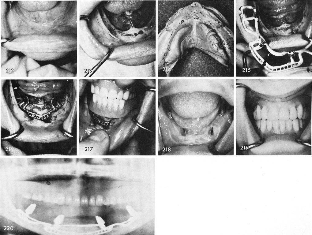
An extremely resorbed clinical picture, fig. 212, reveals also an extremely atrophied mandible, fig. 213. An impression and bite of heavy silicone material was used, fig. 214, which began the processing of the subperiosteal implant, fig. 215, which fitted with extreme accuracy, fig. 216. The temporary denture was carefully checked, fig. 217. The tissues healed remarkably well, fig. 218, and the dentures were completed, fig. 219. Fig. 220 shows the x-ray.
271
|
|
Page 271 |
Next Page |
|
Copyright warning: This information is presented here for free for anyone to study online. We own exclusive internet copyrights on all content presented on this website. We use sophisticated technology to identify and legally close down websites that reproduce copyrighted content without permission - so please don’t do it.
|