| Mandibular Implants (published 1977) | Dr. Leonard I. Linkow |
|
|
Next Page |
tains four, 360° circumferential clasps that can be adjusted over the posts of the subperiosteal implant, figs. 9, 10. A post-operative x-ray shows the accurate fit, fig. 11.
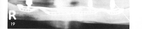
Even with extremely concave situations as seen clinically, fig. 12, the rubber base impression reveals good bone height, fig. 13, for a properly designed implant, fig. 14, to fit with great accuracy and retention over the atrophied mandibular bone, fig. 15. Very often, in order to obtain healing such as was obtained in this case accessory labial and buccal incisions had to be made to the periosteum. Some of the scar tissue seen here is from the accessory incisions, fig. 16. The implant denture is implant borne and does not touch the tissues, figs. 17, 18. The post-operative x-ray, fig. 19.
253

|
|
Page 253 |
Next Page |
|
Copyright warning: This information is presented here for free for anyone to study online. We own exclusive internet copyrights on all content presented on this website. We use sophisticated technology to identify and legally close down websites that reproduce copyrighted content without permission - so please don’t do it.
|