| Mandibular Implants (published 1977) | Dr. Leonard I. Linkow |
|
|
Next Page |
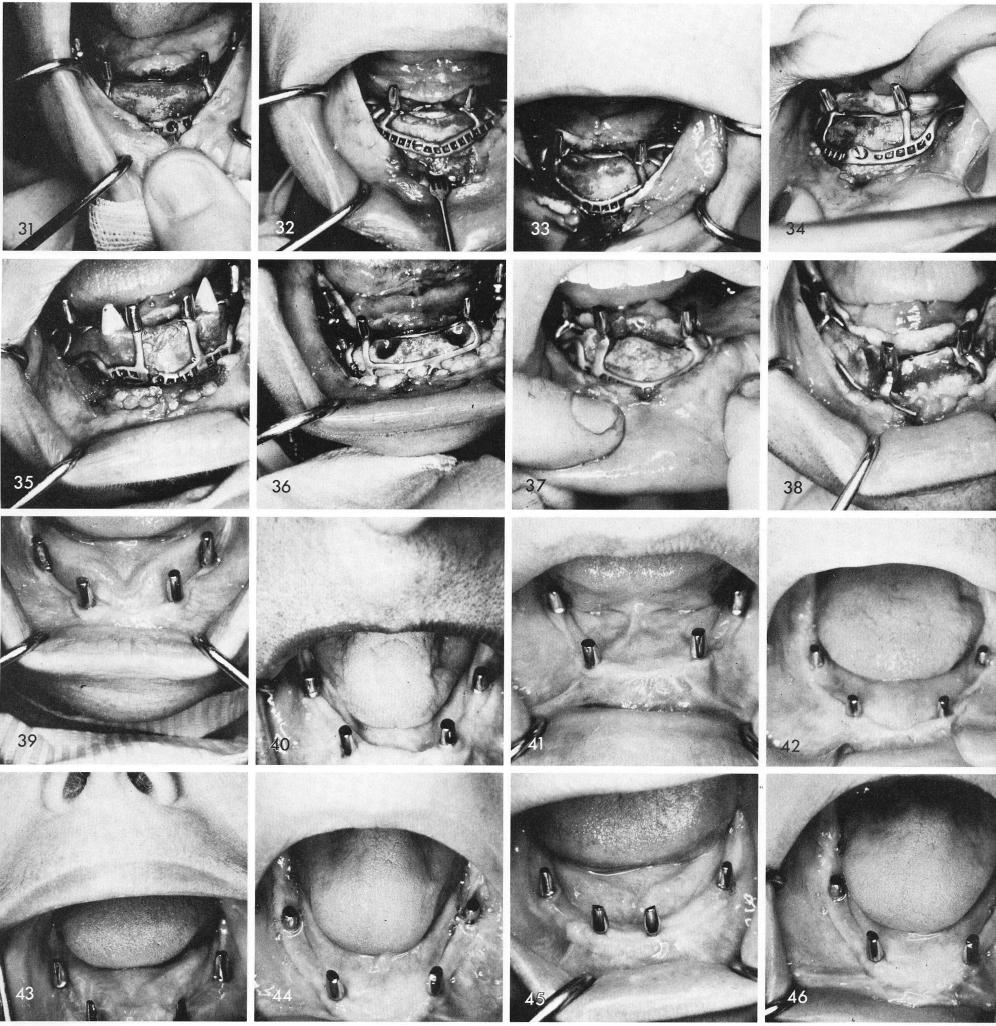
When they fit as exactly as these did and the soft tissues were tenderly cared for and the prosthetic phase carried out with the utmost knowledge and skills, the tissues around the subperiosteal implant posts usually respond remarkably well as seen in figs. 39 through 46. Fig. 47 shows a seventeen and one half year post-operative x-ray of a subperiosteal implant, that was done after an original failure with my first attempt in 1953. Fig. 48 shows the clinical appearance.
250
|
|
Page 250 |
Next Page |
|
Copyright warning: This information is presented here for free for anyone to study online. We own exclusive internet copyrights on all content presented on this website. We use sophisticated technology to identify and legally close down websites that reproduce copyrighted content without permission - so please don’t do it.
|