| Mandibular Implants (published 1977) | Dr. Leonard I. Linkow |
|
|
Next Page |
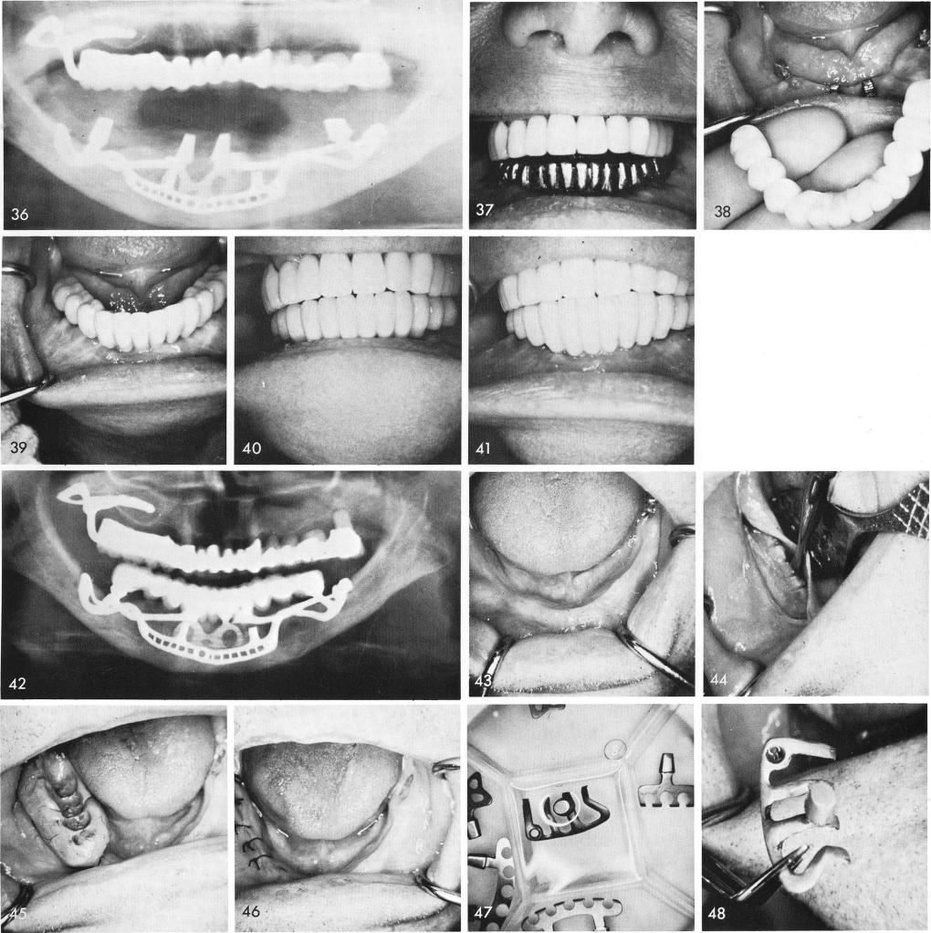
the subperiosteal implant and anterior blade, fig. 36. Castings are fitted, fig. 37, and the completed prosthesis is cemented with hard cement, figs. 38, 39, and the overjet and overbite were still greatly reduced, figs. 40, 41. The post-operative x-ray, fig. 42.
In a totally edentulous situation, fig. 43, it is most important to first prepare the lingual side of the mylohyoid ridge with several shallow grooves and take an impression and bite (Input) for a unilateral subperiosteal implant, figs. 44, 45. The tissues are then closed, fig. 46. At the next visit the sterilized implants, fig. 47, are ready to be fitted and inserted. The unilateral subperiosteal implant must first be tried in, fig. 48, because a full arch temporary splint is much better
216
|
|
Page 216 |
Next Page |
|
Copyright warning: This information is presented here for free for anyone to study online. We own exclusive internet copyrights on all content presented on this website. We use sophisticated technology to identify and legally close down websites that reproduce copyrighted content without permission - so please don’t do it.
|