| Mandibular Implants (published 1977) | Dr. Leonard I. Linkow |
|
|
Next Page |
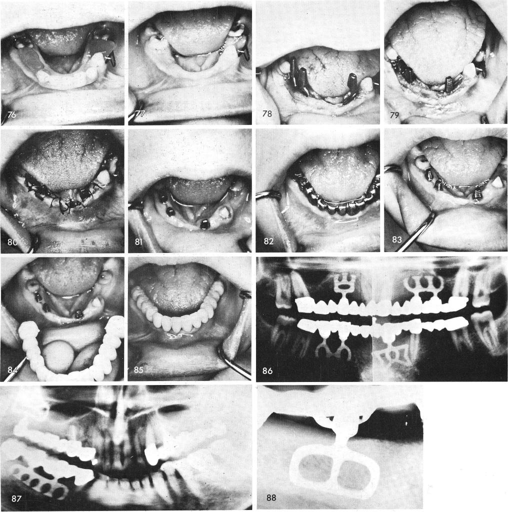
A case of partial anodontia with extremely knife-edged ridges, fig. 76, 77, needed the sup-port of two intra-tooth blades, fig. 78, that were carefully tapped into proper position by first widening the pyramidal shaped knife-edged ridge by removing several milimeters of bone at the crest, thus widening the occlusal table, fig. 79. The tissues were then sutured together, fig. 80. After tissue healing, fig. 81, impressions were taken for individual gold copings, fig. 82. One week later, the case was completed, figs. 83, 84, 85. The post-operative x-ray shows the entire case. It also reveals that the twelve year molars did not have to be involved for added support, fig. 86.
Fig. 87 through 102 shows other cases using intra-tooth bladevents.
188
|
|
Page 188 |
Next Page |
|
Copyright warning: This information is presented here for free for anyone to study online. We own exclusive internet copyrights on all content presented on this website. We use sophisticated technology to identify and legally close down websites that reproduce copyrighted content without permission - so please don’t do it.
|